Fig. 6.
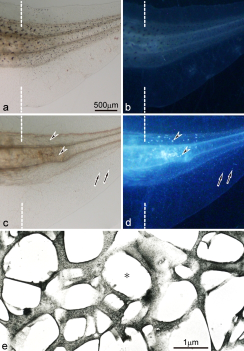
Expression of pigment cells in the 6-day regenerating tail in the wild-type and the mutant (amputated at stage 50). a,b The wild-type regenerating tail observed under transmitted light (a) or incident light (b). c,d The mutant regenerating tail observed under transmitted light (c) or incident light (d). e Ultrastructure of differentiated iridophores in the mutant regenerating tail. Dashed lines indicate the amputation level. Melanophores appeared in the wild-type regenerating tail (a,b), and their distribution was similar to that in the intact tadpole tail. In contrast, white pigment cells (arrows) appeared in the mutant regenerating tail (c,d), and their distribution was similar to that in the intact tadpole tail. A small number of iridophores (arrowheads), which were never present in the intact tadpole tail, appeared in the somites of the mutant regenerating tail in addition to white pigment cells. Differentiated iridophores in the mutant regenerating tail were filled with reflecting platelets, which were irregular in size and shape (asterisk)

