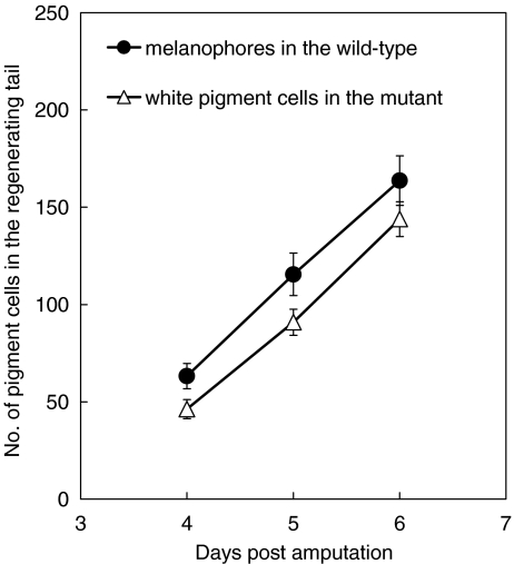
Fig. 7.
The number of newly differentiated pigment cells in the regenerating tail of the wild-type and the mutant. After amputation of the posterior half of the tadpole tail (stage 48/49), melanophores or white pigment cells were counted in the regenerating tail of either the wild-type (n = 15) or the mutant (n = 16), respectively, on days 4, 5, and 6 post-amputation. The number of white pigment cells in the mutant regenerating tail was not statistically different from that of melanophores in the wild-type regenerating tail on days 5 and 6 post amputation (t test, P > 0.05)

