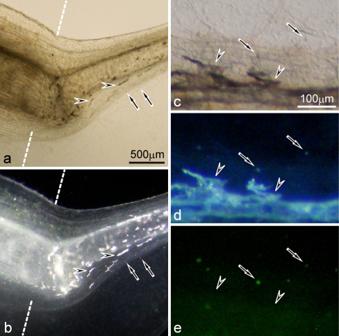
Fig. 9.
Differentiation of iridophores and white pigment cells in the 19-day mutant regenerating tail (amputated at stage 50). a,b The mutant regenerating tail observed under transmitted light (a) or incident light (b). c−e Enlarged view of the mutant regenerating tail of another tadpole observed under transmitted light (c), incident light (d), or blue light (e). Dashed lines indicate the amputation level. White pigment cells (arrows) which were small and aggregated pigment organelles, looked white under incident light and emitted green fluorescence under blue light. In contrast, iridophores (arrowheads) which were large and dispersed pigment organelles, reflected light under incident light, but did not emit green fluorescence under blue light

