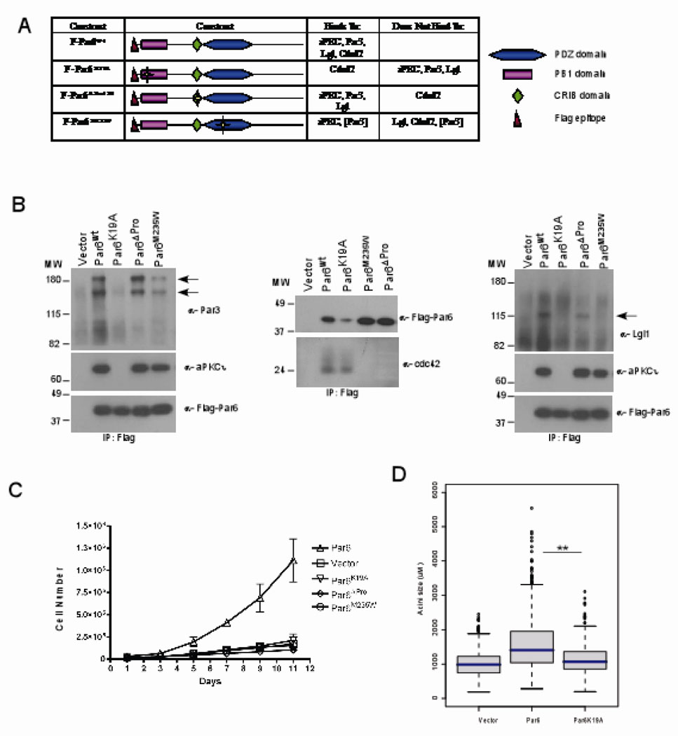
Figure 2. Par6 induced cell proliferation requires interaction with aPKC and Cdc4.
(A) Table summary of the Par6 mutants and their different binding partners. (B) Immunoprecipitaion of cell extracts with Par6 (anti-flag antibodies) and immunoblotted with anti-aPKCi, anti-Flag, anti-Par3, anti-Cdc42 and anti-lgl antibodies. (C) Growth curve of vector control cells compared to Par6wt and mutant Par6 cells over a period of 11 days in EGF free media. Data are means +/− s.d. of estimated cell numbers from three independent experiments. (D)= Distribution of acini size (circumferential area) of Day 12 structures grown in 0.5ng/ml EGF. Each condition represents approximately 800 acini structures from three dependent experiments. The P-value between Vector and Par6wt or Par6wt and K19A is less than 0.0001 calculated by Mann-Whitney test.

