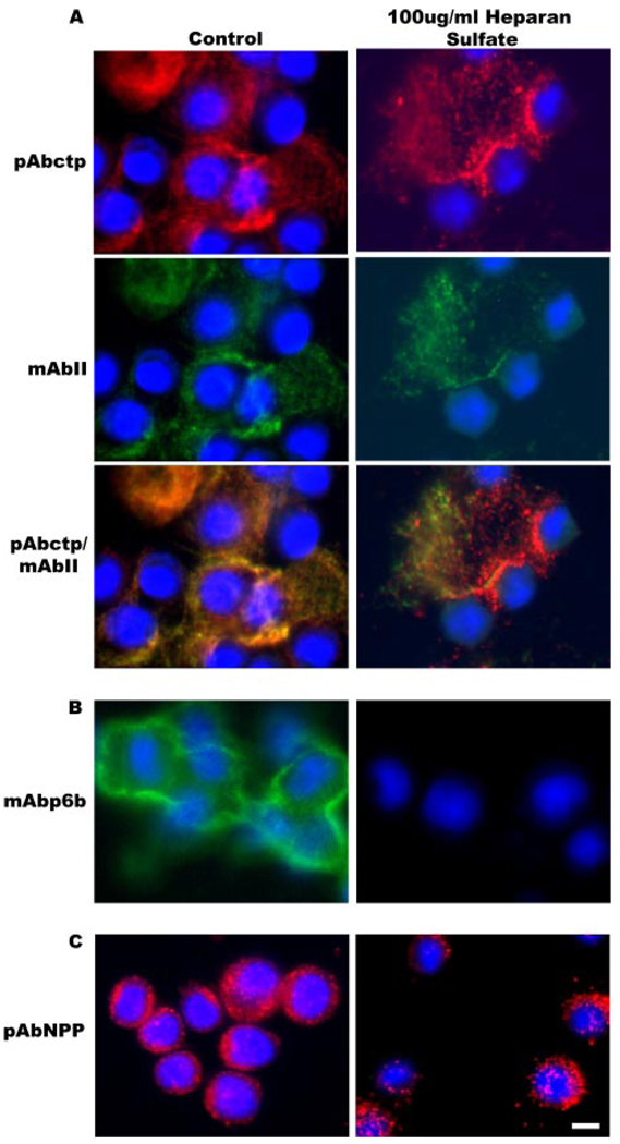
FIGURE 7. Specific localization of Npp[p6b7] within the pericellular matrix.
Cells were maintained in culture for 48 h during which the pericellular matrix was allowed to assemble in the absence or presence of heparan sulfate. A, double label with polyclonal antibody to the carboxyl telopeptide (ctp) of collagen α1(XI), which remains associated with the major triple helix after proteolytic removal of the carboxyl propeptide, and a monoclonal antibody to collagen type II. Localization of collagen type XI and collagen type II are not significantly affected by the presence of heparan sulfate in the growth medium. B, immunolabel with monoclonal antibody to p6b (containing heparan sulfate binding site 2). Note diminished labeling with monoclonal antibody to p6b in the presence of heparan sulfate. C, immunolabel with polyclonal antibody to collagen α1(XI)Npp (containing heparan sulfate binding site 1). Note punctate distribution of the Npp epitope with relatively unaltered localization in the presence of heparan sulfate. Nuclei were stained with DAPI (blue). Scale = 10 µm.

