
Figure 6.
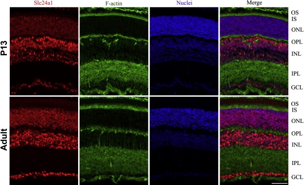
Immunolocalization of Slc24a1 Expression in P13 and Adult Mouse Retinal Layers
The slides were stained for Slc24a1 with goat anti-rabbit IgG conjugated with AlexaFluor 594 (red), F-actin using AlexaFluor488 conjugated phalloidin (green), and nuclei using Hoechst 33342 (blue). Merge represents an overlay of images from the first three columns in P13 and adult mouse retinal layers, respectively. The localization pattern illustrates a strong signal in the inner segment, outer and inner nuclear layers, and ganglion cell layer. The scale bar represents 50 um. OS, outer segment; IS, inner segment; ONL, outer nuclear layer; OPL, outer plexiform layer; IPL, inner plexiform layer; GCL, ganglion cell layer.