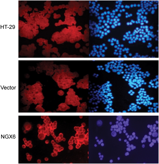
Figure 6.
β-Catenin is relocated to cell membrane caused by NGX6 HT-29, pcDNA3.1(+)/HT-29, and pcDNA3.1(+)/NGX6/HT-29 cells were cultured in multichamber slides. After incubation, cells were fixed and then detected for β-Catenin expression by Cy3-labeled immunofluorescence staining. DAPI-stained cells were used as an internal control. It showed that β-catenin protein localizes mainly on cell membranes in pcDNA3.1(+)/NGX6/HT-29 cells, and low-level staining observed in cytoplasm and nucleus. β-Catenin proteins present diffuse staining throughout cytoplasm, nucleus, and cell membrane in HT-29 and pcDNA3.1(+)/HT-29 cells.

