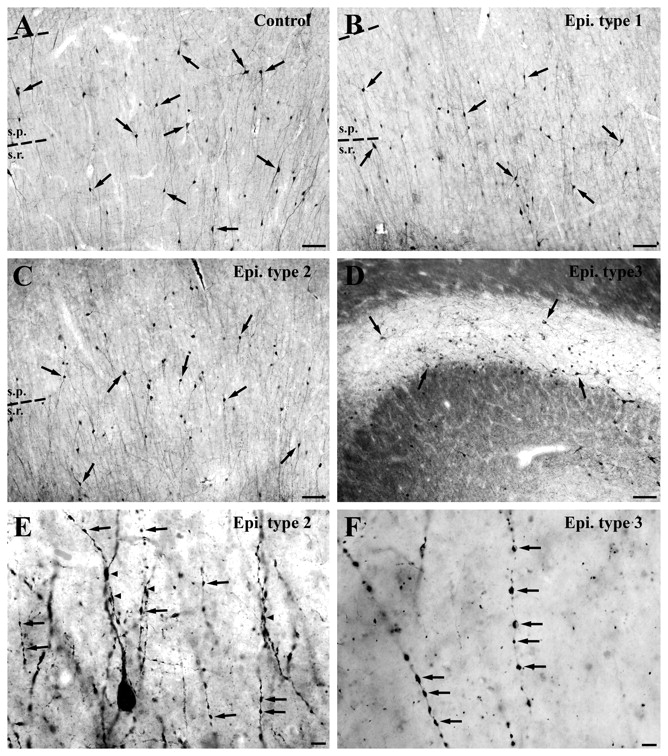
Figure 2.

Distribution and morphology of calretinin-positive cells in the human control and epileptic CA1 region. (A) Numerous immunoreactive cells with smooth dendrites can be seen in all layers of the CA1 region in control samples with short post-mortem delay. (B and C) The number of immunolabelled cells and the extent of their dendritic trees are quite similar in the non-sclerotic Type 1 (B) and Type 2 (C) samples compared to controls, although in the latter cases the dendrites are varicose and segmented. (D) The number of calretinin-positive cells is decreased significantly in the sclerotic epileptic cases. The area of the sclerotic hippocampi is reduced because of the shrinkage of the CA1 region caused by the loss of CA1 pyramidal cells. (E and F) High-power light micrographs show the morphological alterations of the calretinin-positive dendrites in epileptic cases. In the non-sclerotic Type 2 samples (E), almost all of them show signs of degeneration, the dendrites have become beaded and segmented (arrows, E). Dendro-dendritic connections are less frequent than in controls (arrowheads, E). In sclerotic cases (F) only fragmented dendrites can be seen, most of the beads are separated (arrows, F). Scale bars: A–D = 100 µm; E and F = 20 µm; s.p. = stratum pyramidale; s.r. = stratum radiatum.
