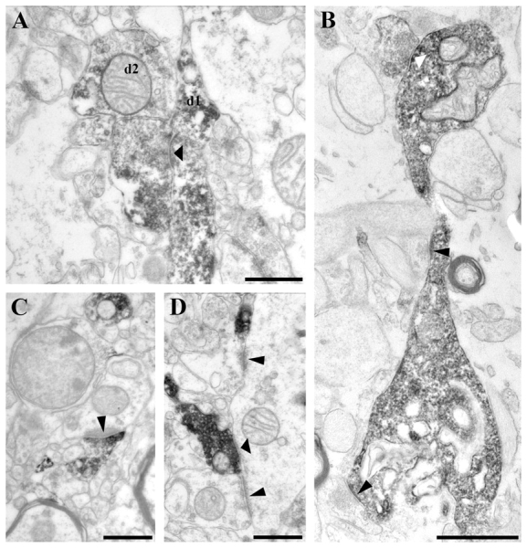
Figure 4.

High-power electron micrographs from control (A, C and D) and epileptic (B) CA1 region. Terminals of calretinin-positive interneurons establish symmetric synaptic contacts (A, B and D, thick arrowheads). They frequently terminate on other calretinin-positive interneuron dendrites (A; d1). Dendrites of unlabelled interneurons also receive synaptic contacts from calretinin-positive inhibitory terminals (D). At the border of stratum lacunosum-moleculare a part of the calretinin-positive terminals establish asymmetric synaptic contacts with very thick post-synaptic density most frequently targeting unlabelled dendrites (C, thin arrowheads). B shows a varicose dendrite surrounded by glial processes. The beads are degenerating, although they still receive synaptic input, a symmetric synaptic contact from a calretinin-immunopositive bouton (thick white arrowhead) and two asymmetric synaptic contacts from unlabelled terminals (thin black arrowheads). Scale bars: A, C and D = 0.5 µm; B = 10 µm.
