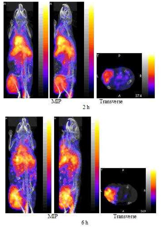
Fig. 5.

PET and fused CT projections obtained by imaging one mouse bearing a SUM190 tumor in the left thigh 2 h (top row) and again at 6 h (bottom row) post injection of 100 μL (0.22 MBq) of 18F labeled nanoparticle with each acquisition requiring 30 min. Each presents an anterior (left panel) and left lateral projection (middle panel), both Maximum Intensity Projections (MIP) and a transverse slice (right panel) of the acquisition centered on the tumor.
