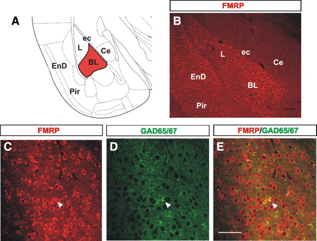
Figure 1.
FMRP is expressed in the majority of interneurons in the BL. A, Schematic of a coronal view of a brain at the level of the amygdala with the BL highlighted in red. B, Image of FMRP immunostaining at P21 showing the intense and high levels of FMRP expression in the L and BL in contrast to the much lesser immunostaining in other regions of the amygdala such as the central nucleus. C–E, The majority of GAD65/67-positive neurons also show FMRP expression in the BL at P21 (arrowheads). Ce, Central nucleus of the amygdala; ec, external capsule; EnD, endopiriform nucleus; L, lateral nucleus of the amygdala; Pir, piriform cortex. Scale bars: B, E (for C–E), 10 μm.

