Figure 3.

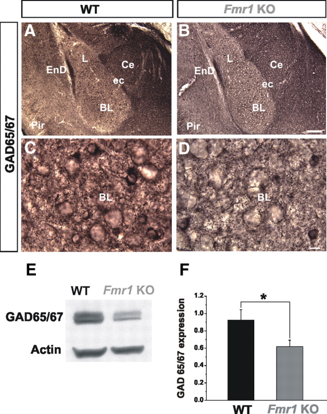
GAD65/67 levels are reduced in Fmr1 KOs. A, B, Normal gross morphology and positioning of GAD65/67+ inhibitory neurons in Fmr1 KO mice (B) compared with WT (A) in the BL is shown at P21 at low-power magnification. C, D, Qualitative decrease in GAD65/67 immunostaining is observed in Fmr1 KOs compared with WT as shown at high-power magnification. E, F, Western blot analyses reveal that GAD65/67 expression levels are significantly decreased in the Fmr1 KOs, compared with WT in the BL. BL, Basolateral nucleus of the amygdala; Ce, central nucleus of the amygdala; ec, external capsule; EnD, endopiriform nucleus; L, lateral nucleus of the amygdala; Pir, piriform cortex. *p < 0.05. Scale bars: B, 100 μm; D, 10 μm.
