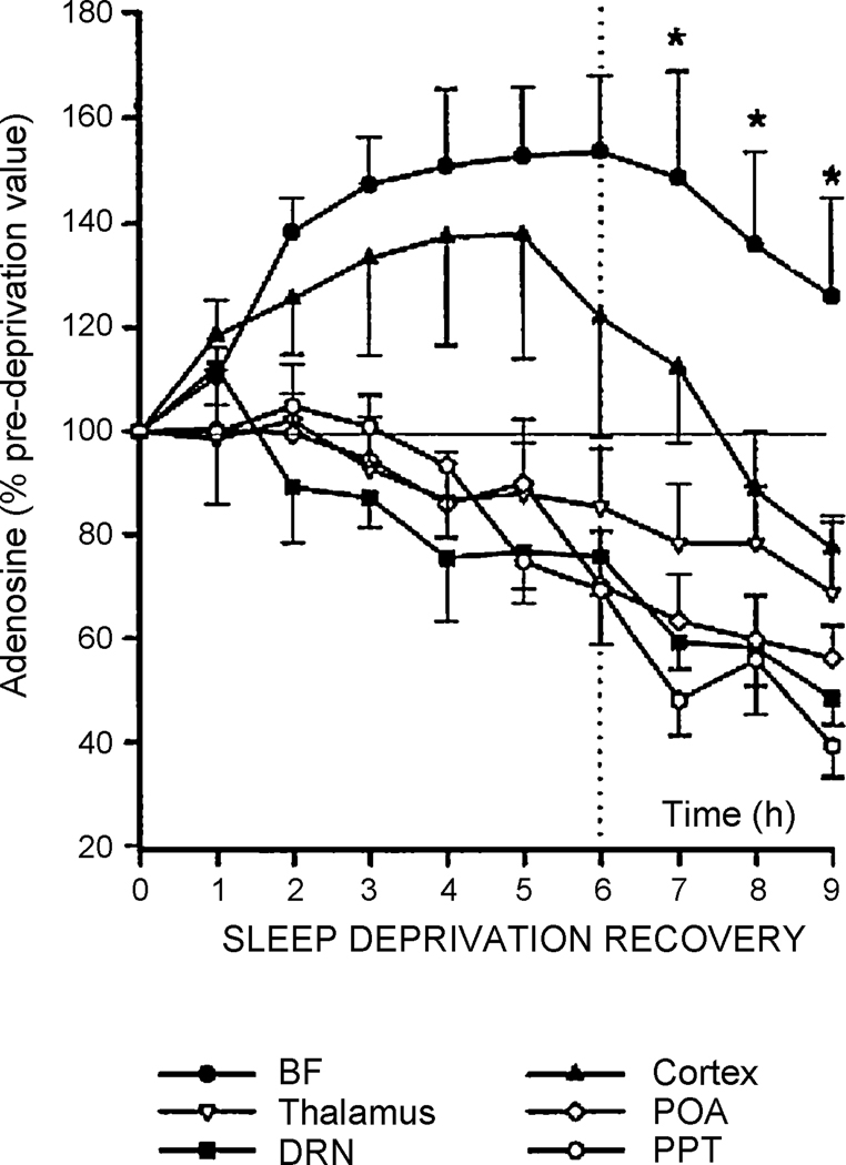
Figure 2.
Alterations in extracellular adenosine measured by microdialysis following sleep deprivation and in subsequent recovery sleep in cats. Cats were sleep deprived for 6 hours and then allowed to sleep for 3 hours. Extracellular adenosine was measured at the beginning of the experiment by microdialysis and then in 1 hour intervals for the duration of the experiment. Adenosine is presented as a percentage of the baseline value. Extracellular adenosine increases during sleep deprivation only in the basal forebrain and cortex. In the other areas studied, adenosine levels progressively decline. Adenosine stays elevated during recovery sleep only in the basal forebrain. BF, basal forebrain; POA, preoptic area of the hypothalamus; DRN, dorsal raphe nucleus; PPT, pedunculopontine tegmental area. (Porkka-Heiskanen et al. 2000; reprinted with permission).

