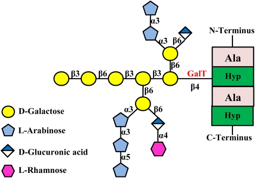
Figure 1.
AG structure of an AGP molecule. The protein backbone containing a clustered noncontiguous Hyp motif is shown. Although the two Hyp residues in the protein backbone are both glycosylated with AG when expressed in tobacco cells, only one AG side chain is shown here for simplicity. The focus of this study is the GalT enzyme (shown in red) that adds the first Gal residue onto the AGP peptide backbone. Monosaccharide symbols used here are based on the Symbol and Text Nomenclature for Representation of Glycan Structure as proposed by the Consortium for Functional Glycomics (http://glycomics.scripps.edu/CFGnomenclature.pdf). Modified from Tan et al. (2004).

