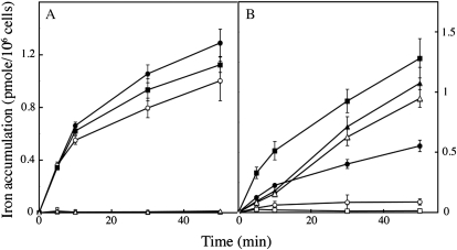
Figure 4.
Nonreductive iron uptake in C. velia compared to reductive and siderophore-mediated iron uptake in S. cerevisiae. C. velia (A) and S. cerevisiae (B) cells were grown in iron-deficient medium and treated as described in Figure 2. Iron (1 μm) was added to the cell suspensions, in the form of 55Fe(III)-citrate (1:20; circles), 55Fe(II)-ascorbate (1:10; squares), or 55Fe-ferrichrome (triangles), with (white symbols) or without (black symbols) addition of the ferrous chelator BPS (100 μm). Aliquots were taken at intervals and washed three times with Mf medium containing EDTA, BPS, and desferrichrome (10 mm each) before counting. Means ± sd from three experiments are shown.

