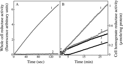
Figure 5.
Whole cell and intracellular reductase activity. C. velia and S. cerevisiae cells were grown in iron-deficient medium and treated as described for Figure 2. A, Trans-plasma membrane electron transfer for whole cells was monitored by fluorimetric analysis of the formation of resorufin from resazurin (10 μm). Both C. velia (curve 2) and S. cerevisiae, used as a control (curve 1), were analyzed at a concentration 100 × 106 cells/mL. B, NAD(P)H-dependent ferrireductase activity of soluble whole-cell homogenate of C. velia. Cells were grown in iron-rich (1 μm; black symbols) or iron-limited (10 nm; white symbols) Mf medium, washed, and broken up with glass beads. The soluble fraction (10,000 g supernatant) was tested for ferrireductase activity by measuring absorbency at 535 nm after the addition of 0.5 mm Fe(III)-citrate (1:20), 1 mm BPS, and either 1 mm NAPDH (curves 1 and 3) or NADH (curves 2 and 4) as the electron donor. One representative experiment out of three (A) or two (B) is shown.

