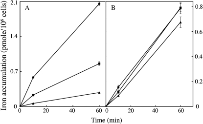
Figure 6.
Effect of Fe3+ ligand concentration on iron accumulation. C. velia (A) and S. cerevisiae (B) cells were grown in iron-deficient medium and treated as described for Figure 2. Iron (1 μm) was added to the cell suspensions, in the form of 55Fe(III)-citrate, at different Fe(III):citrate ratios. Circles: 10 μm citrate [Fe(III)-citrate 1:10]; squares: 50 μm citrate [Fe(III)-citrate 1:50]; triangles: 250 μm citrate [Fe(III)-citrate 1:250]. Aliquots were taken at intervals and washed three times with Mf medium containing EDTA, BPS, and desferrichrome (10 mm each) before counting. Means ± sd from three experiments are shown.

