Figure 5.
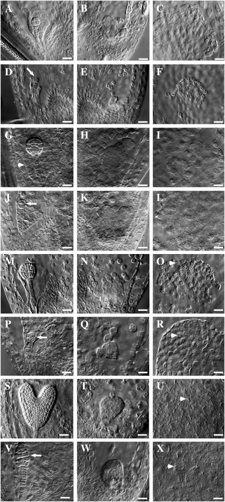
Embryo and endosperm development in wild-type and ctf7+/2 siliques. Wild-type (A–C, G–I, M–O, and S–U) and ctf7+/2 (D–F, J–L, P–R, and V–X) embryo and endosperm whole-mount images are shown. A, Wild-type octant-stage embryo. The MCE is still at the NCD stage. B, CZE of the embryo sac in A. C, PEN of the embryo sac in A. D, ctf7 embryo showing aberrant cell division. The arrow indicates the division plane. E, CZE of the embryo sac in D. F, PEN of the embryo sac in D. G, Wild-type globular embryo. The MCE shows signs of cellularization (arrowhead). H, CZE of the embryo sac in G. Endosperm nodules and chalazal cyst are visible. I, PEN of the embryo sac in G. J, ctf7 embryo showing aberrant cell division. The arrow indicates the division plane. K, CZE of the embryo sac in J. L, PEN of the embryo sac in J. M, Wild-type early-heart-stage embryo. N, CZE of the embryo sac in M. O, PEN of the embryo sac in M. The arrowhead indicates NCDs starting to cellularize. P, ctf7 embryo showing aberrant cell division. The arrow indicates abnormal cell shape and cell organization. Q, CZE of the embryo sac in P. R, PEN of the embryo sac in P. The arrowhead indicates NCDs starting to cellularize. S, Wild-type torpedo-stage embryo. T, CZE of the embryo sac in S. NCDs are still present. U, PEN of the embryo sac in S. The arrowhead shows cells that have completed cellularization. V, ctf7 embryo with an abnormal shape and disorganized cell order. The arrow indicates abnormal cell organization. W, CZE of the embryo sac in V. X, PEN of the embryo sac in V. The arrowhead shows cells that have completed cellularization. Bars = 10 μm.

