Figure 4.
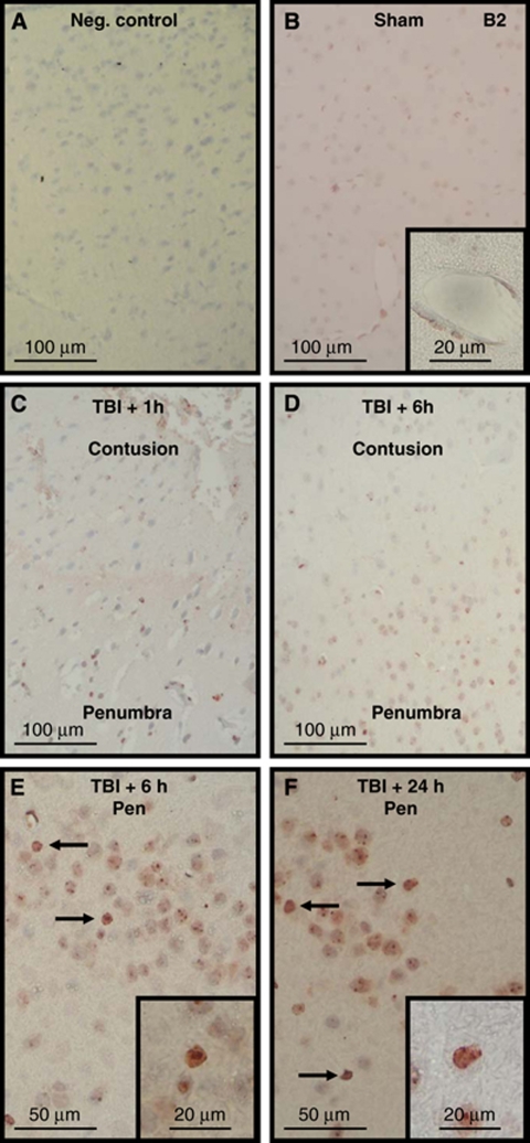
Immunohistochemistry for kinin B2 receptors in sham-operated and traumatized C57/BL6 mice. Negative control by omission of the primary antibody (A, negative control). Staining was performed in sham-operated mice (B) or in mice killed 1 (C), 6 (D, E), or 24 h (F) after experimental TBI (C57/BL6; n=4 per group). In contrast to the B1 receptor the B2 receptor is also found in cerebrovascular vessels (B, inset). Again, no staining was observed within the contused tissue (C, D), whereas cellular staining (arrows) became apparent in the traumatic penumbra with time (C to F).

