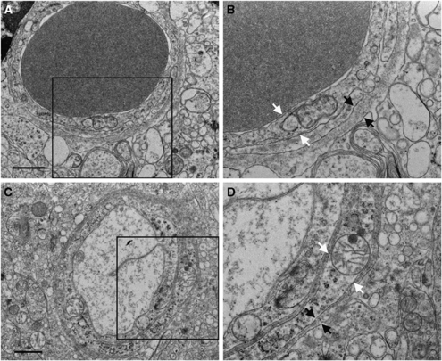
Figure 3.
Electron microscopy of cortical microvessels at postnatal day 21. Microvessels from PTU-treated animals (A and B) and control animals (C and D) appear to have normal endothelial cell (white arrows) and basal membrane structure (black arrows). Panels B and D are enlargements of the framed areas in panels A and C, respectively. Scale bar in panels A and C=1 μm.

