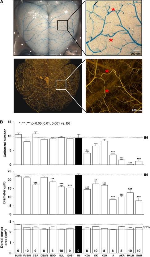
Figure 1.
Wide genetic variance of the native pial collateral circulation exists among 15 inbred mouse strains. (A) Mouse pial arterial circulation. Upper panels show vasculature after clearing, dilation, fixation, and filling with high-viscosity polyurethane restricted from capillary transit. Measurements in (B) and Figures 2, 3, 4, 5, 6 to 7 and Supplementary Figures are for collaterals interconnecting the middle and anterior cerebral artery trees (*). Lower panels show high-viscosity microfil (Flow Tech Inc), followed by optical clearance with methylsalicylate, to highlight penetrating arterioles and confirm that native collaterals are confined to the pial surface. An occasional center-most collateral segment between two penetrating arterioles that escapes filling is easily identified as part of a single collateral (lower enlargement). (B) Variation in collateral number and diameter (per hemisphere) is unrelated to small variation in cortex area (or body weight, Supplementary Figure 1). B6, C57BL/6; number of animals given at base of columns also applies to subsequent figures, unless indicated otherwise.

