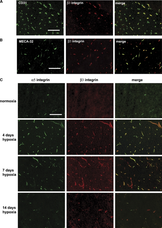
Figure 2.
Expression of the β3 integrin on angiogenic capillaries in the hypoxic central nervous system (CNS). Dual-immunofluorescence (IF) was performed on frozen sections of cerebral cortex from normoxic or hypoxia-exposed mice using monoclonal antibodies specific for: (A) CD31 (Alexa Fluor 488) or β3 integrin (Cy-3), (B) MECA-32 (Alexa Fluor 488) or β3 integrin (Cy-3), and (C) α5 integrin (Alexa Fluor 488) or β3 integrin (Cy-3). Scale bar=50 μm. Note that after 4 days hypoxia, only a fraction of the CD31-positive capillaries stained positive for β3 integrin, but all MECA-32-positive capillaries stained positive for β3 integrin, confirming that β3 integrin is expressed by angiogenic endothelial cells within the hypoxic CNS. Also note that hypoxia promoted strong induction of both α5 and β3 integrins on capillaries, though the timing of expression of the two subunits was different; α5 integrin expression peaked at 4 days hypoxia, whereas β3 expression peaked at 7 days hypoxia.

