Figure 3.
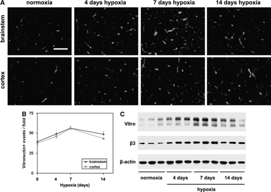
Hypoxic induction of vitronectin on cerebral blood vessels. (A) Frozen sections of brainstem or cerebral cortex taken from mice exposed to normoxia or 4, 7, or 14 days hypoxia were immunostained for vitronectin. Scale bar=50 μm. (B) Quantification of vitronectin-positive vessels. Experiments were performed with three different animals per condition, and the results expressed as the mean±s.e.m. of the number of capillaries positive for each antigen per field of view. (C). Western blotting confirmation of hypoxic induction of β3 integrin and vitronectin in brain lysates. Note that cerebral hypoxia promoted increased expression of vitronectin and the β3 integrin subunit, with the greatest effect after 7 days hypoxia.

