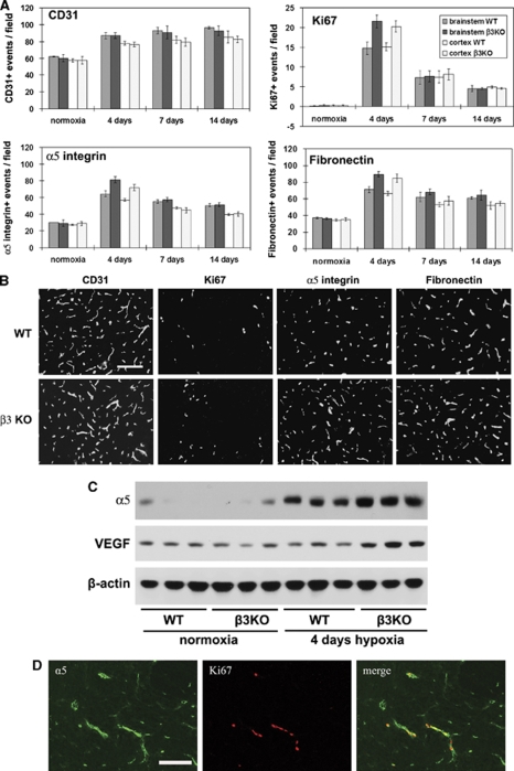
Figure 6.
Comparison of the hypoxic-induced angiogenic response, in the wild-type and β3 integrin-null central nervous system (CNS). β3 integrin-null and wild-type littermate control mice were maintained at normoxia or exposed to 8% hypoxia for 4, 7, or 14 days before frozen brain sections immunostained to assess blood vessel density (CD31), cell proliferation (Ki67), and expression of α5 integrin and fibronectin. (A) Quantification of analysis. Brain sections were examined for the number of vessels that stained positive for the different antigens. Experiments were performed with three different animals per condition, and the results expressed as the mean±s.e.m. of the number of capillaries positive for each antigen per field of view. (B) Representative images of immunofluorescence (IF) of brainstem after 4 days hypoxia. Scale bar=50 μm. Note that cerebral hypoxia promoted an increased capillary density (CD31) in the brains of wild-type and β3 integrin-null mice, with no significant differences observed between the two groups at any time point. In contrast, compared with wild-type mice, after 4 days hypoxia, brain endothelial cells (BEC) in β3 integrin-null mice showed a significantly increased mitotic index (Ki67-positive events), with parallel increases in the number of vessels positive for α5 integrin and fibronectin. (C) Western blot analysis. Note that after 4 days hypoxia, levels of the α5 integrin and vascular endothelial growth factor (VEGF) were significantly higher in the β3 integrin-null mice compared with wild-type controls (n=3 mice per condition). (D) Colocalization of α5 integrin and the proliferation marker Ki67 in the 4-day hypoxic CNS. Scale bar=50 μm.

