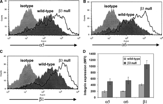
Figure 7.
Compensatory upregulation of β1 integrin expression on β3 integrin-null brain endothelial cells (BEC). Wild-type or β3 integrin-null BEC were cultured on fibronectin. On reaching confluence, flow cytometry was used to quantify BEC expression of the integrin subunits α5 (A), α6 (B), or β1 (C). (D) Quantification of expression of β1 integrins by β3 integrin-null BEC. All points represent the mean±s.e.m. of the mean fluorescent intensity (MFI) of three separate experiments. Note that relative to wild-type cells, β3 integrin-null BEC showed a marked upregulation of the α5, α6, and β1 integrin subunits.

