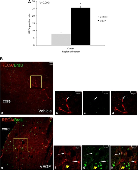
Figure 5.
Vascular endothelial growth factor (VEGF) increases angiogenesis after traumatic brain injury (TBI). Animals were killed 90 days after TBI and the number of blood vessels was counted in the perilesion cortex. Significantly more vessels were seen in the cortical area surrounding the damage in VEGF-treated mice (A). (B) A photomicrograph ( × 200) showing that many of the blood vessels in this area had BrdU+ cells colocalizing with the endothelial marker RECA1 (white arrows) in vehicle- (a–d) and VEGF (e–h)-treated animals, but the number of vessels with colocalization was significantly larger in VEGF-treated animals. Note that in VEGF-treated mice, 5-bromo-2-deoxyuridine (BrdU)/RECA1 double-positive cells (white arrows) have a different morphology from that of BrdU+ RECA1 cells (yellow arrow).

