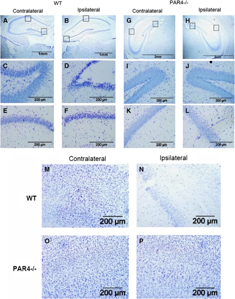
Figure 2.
Neurodegeneration in the ipsilateral, but not contralateral hippocampus and cortex of wild-type but not PAR4 null mice after I/R injury. Representative photographs with cresyl violet staining at low (A, B, G, H) and high (C to F and I to P) magnification of the wild-type (n=5) ipsilateral hippocampus (B, D, and F) and cortex (N); wild-type (n=5) contralateral hippocampus (A, C, and E) and cortex (M); PAR4−/− ipsilateral hippocampus (H, J, and L) and cortex (P); PAR4−/− contralateral hippocampus (G, I, and K) and cortex (O).

