Figure 1.
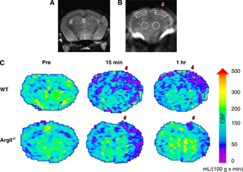
Regions of interest (ROIs) for cerebral blood flow (CBF) calculations. (A–C) Regions of interest and CBF maps for arterial spin labeling (ASL) MRI mouse brain scans. (A) Coronal brain slice used for CBF measurements. (B) Six ROIs were used to determine CBF, three from each hemisphere. Regions contralateral and ipsilateral to the traumatic brain injury (TBI) (indicated by the red arrow) were chosen in the cortex, subcortex, and striatum. (C) Cerebral blood flow map of an arginase II-deficient (ArgII−/−) mouse and a wild-type (WT) mouse before TBI, 15 minutes after TBI, and 1 hour after TBI. The ArgII−/− mice show less CBF blunting and/or better CBF recovery. Maps were created by applying the relative CBF (rCBF) equation (rCBF = λ × 60,000 × ((1/T1(selective))−(1/T1(nonselective))) to T1 maps of the brain created from the ASL scans. A color map was applied. Relative CBF units: mL/100 g/minute. Red arrows indicate the site of TBI impact.

