Figure 2.
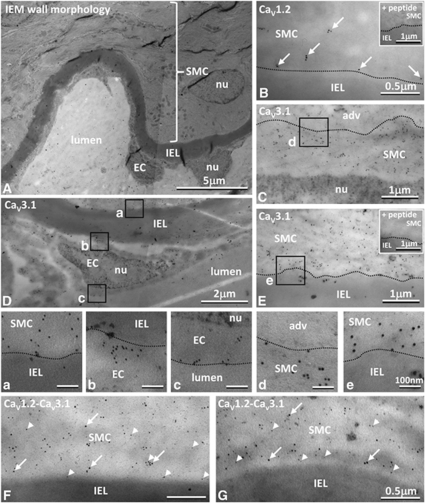
Location of L- and T-type channels at the ultrastructural level. (A) Tissue prepared for immunoelectron microscopy shows normal vessel morphology with smooth muscle cells (SMCs) and endothelial cells (ECs) separated by the internal elastic lamina (IEL); with myosin filaments oriented to the longitudinal cell axis and accumulations of mitochondria within the cells. (B–G) Gold labelled CaV1.2 (10 nm) and CaV3.1 (5 nm) were found in the SMC cytoplasm and at cell membranes (B, C, E–G). CaV3.1 was also found in ECs (D). Boxes in C–E shown at higher magnification in panels a–e. Gold particles were rarely found over nuclei (nu: C, D, panel c), adventitia (adv: C, panel d), IEL (B, E, panels a, b, and e, F, G), or lumen (panel c), and staining was abolished after incubation with appropriate corresponding immunogenic peptide (insets in B, E).

