Figure 6.
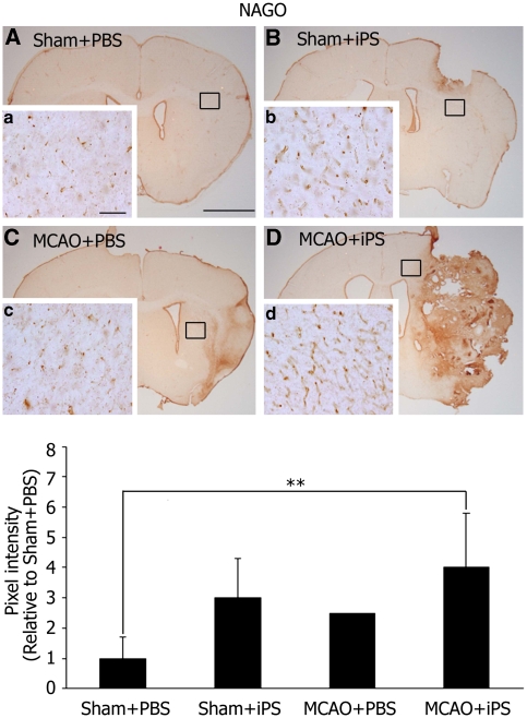
NAGO was abundantly expressed outside of tumors. NAGO, a protein marker of vascular endothelial cells, was observed at basal level at 28 day after the iPS cell transplantation in the Sham+PBS group (A), which was highly induced in the periphery of tumors in the Sham+iPS (B), and slightly induced in the MCAO+PBS groups (C). Such NAGO induction was more striking in the MCAO+iPS group (D); (a to d) represent magnification of the boxed areas in (A, B, C, D), respectively. Scale bar=2 mm (A), 100 μm (a). The intensity of NAGO staining was significantly higher in the MCAO+iPS group compared with Sham+PBS group (**P<0.01). Statistical analysis: nonrepeated measures analysis of variance (ANOVA) and Bonferroni correction. Data represent mean±s.d. iPS, induced pluripotent stem; MCAO, middle cerebral artery occlusion; NAGO, N-acetylglucosamine oligomer; PBS, phosphate-buffered saline.

