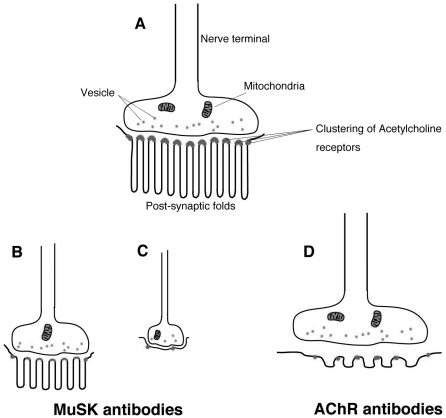
Figure 2.
Schematic appearances of NMJs observed in normal volunteers and patients. A: Normal NMJ. AChRs are concentrated at the peaks of abundant, well-preserved and intricately twisted junctional folds. B and C: NMJ in EAMG induced by MuSK antibodies, CMS with MuSK or Dok-7 mutations. Small NMJ in both pre- and post-synaptic structures. (B) Attenuation of AChR and reduced twisting of synaptic folds at the post-synaptic membrane without widened synaptic space. (C) Disappearance of post-synaptic folds with preserved synaptic space. D: NMJ in MG patients with AChR antibodies. Complement-mediated lysis of post-synaptic membranes. The myasthenic junction has a reduced number of AChR, simplified synaptic folds and a widened synaptic space with a normal nerve terminal.

