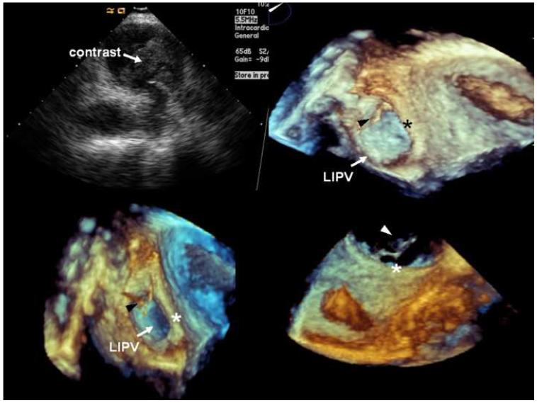
Figure 2.
Echocardiographic monitoring of contrast injection in the VOM. A, Intracardiac echocardiographic snapshot of the left pulmonary veins, showing contrast appearance in the carina. B, C, and D, Three-dimensional transesophageal echocardiography snapshots focusing in the lateral wall of the LA, left lateral ridge (*), and left inferior pulmonary vein (LIPV). The first appearance of echocardiographic contrast agent (arrowheads) was detected in the lateral ridge, anterior to the left inferior pulmonary vein.

