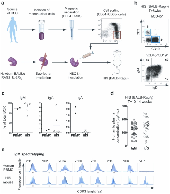
Figure 1. Characterization of B cells in naïve HIS mice.
(A) HIS mice are generated by transplanting sorted human CD34+CD38− HSC-enriched cell fraction into conditioned (3.5 Gy irradiation) newborn Rag2−/− IL-2Rγc−/− mice. After 6 to 8 weeks post-transplantation, human lymphoid and myeloid cell subsets are found in the blood and lymphoid organs of the reconstituted animals. (B) The top panel shows the flow cytometry analysis of human T (CD3+) and B (CD19+) cells found in the spleen of 8-week old HIS mice. The expression of IgM and IgD on the surface of B cells is depicted on the low panel. (C) The relative proportion of B cells expressing an IgM, IgG or IgA BCR was determined by quantitative PCR for both control PBMC and HIS splenocytes. Percentages represent the frequency of IgM, IgG and IgA (CHμ, CHγ and CHα) containing VH PCR products out of the total VH PCR products from each spleen. The horizontal bars indicate the mean value. (D) The graph shows the concentration of total IgM and IgG (mean values: horizontal bars) in the plasma of 10- to 14-week old naive HIS mice. (E) The naive IgM B cell repertoire of HIS mice was evaluated on splenocytes by performing a BCR immunoscope for each VH family. The profiles obtained with control human PBMC are also shown.

