
Figure 2. Comparison of Luteolin Effects on Gene Expression in PC-3 and DU-145 Human Prostate Cancer Cells.
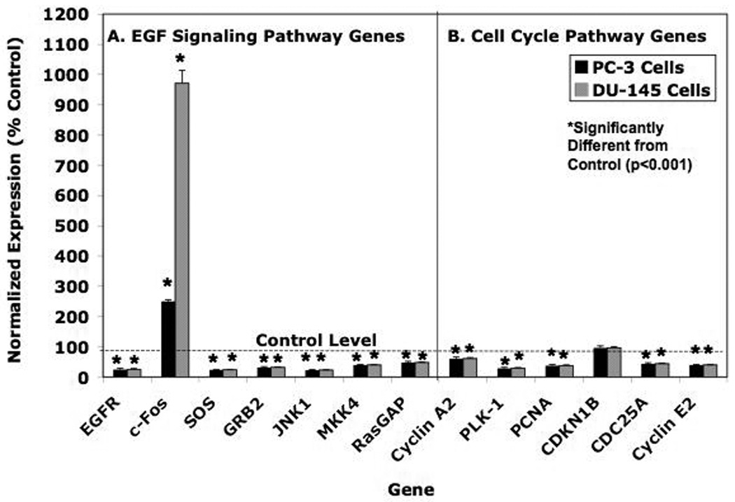
PC-3 and DU-145 cells were grown as described in Material and Methods in either DMEM-F12 Media (PC-3 cells) or Minimal Essential Medium (MEM) supplemented with 10% Fetal Calf Serum (FCS). Twenty-four hours following plating, the cells were treated for 6 hours with with 17.5 µM luteolin (added in EtOH). RNA prepared from these cells was analyzed by real-time PCR (qPCR). Relative expression values are the mean ± SEM for three independent RNA sets normalized to 18S RNA as described in detail [30] presented as % control where the EtOH controls represent 100%. Panel A = EGF Signaling Pathway Genes. Panel B = Cell Cycle Pathway Genes. Data from three replicate experiments were analyzed statistically by ANOVA with Tukey’s Test on the means with Instat Software as described in Methods.