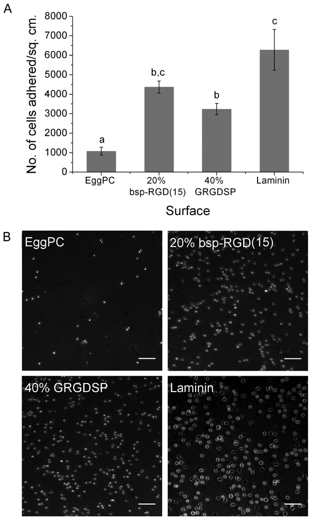
Figure 3.
Neural stem cell adhesion on surfaces after 2 h incubation. (A) Number of cells adhered per unit area, as determined by counting DAPI-stained nuclei. Bars show mean ± SEM (n ≥ 6). Adhesion on 20% bsp-RGD(15) and 40% GRGDSP surfaces was significantly greater than EggPC control; with adhesion on 20% bsp-RGD(15) being comparable to that on laminin. (B) Phase contrast images of adhered cells. Scale bar: 100 µm.

