
Figure 1.
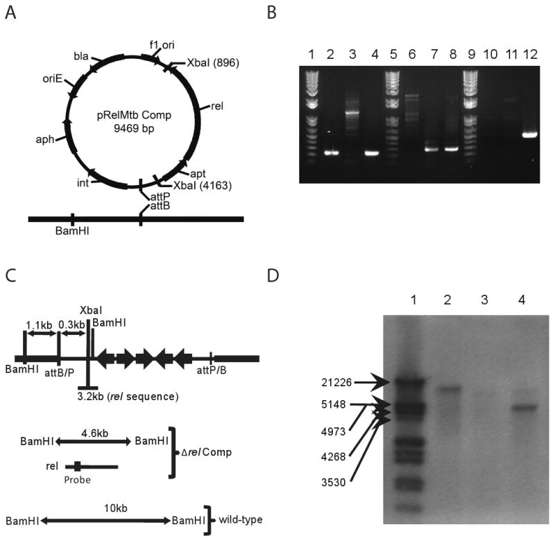
Complementation of Δrel. A. Diagram of expected recombination between integrating plasmid and genomic DNA. B. PCR analysis of genomic DNA. Lanes 2-4 show the presence of rel in wild-type and rel Comp strains. Lanes 6-8 show the presence of a region of the hygromycin gene marking the deletion of rel in both the Δrel and rel Comp strains. Lanes 10-12 show the presence of a region of the kanamycin gene marking the complementation of rel in only the rel Comp strain. C. Diagram of expected sizes of genomic gene fragments expected to bind to probe recognizing region of rel coding sequence. D. Southern blot showing rel as a single copy gene present in the correct fragment size in both wild-type and rel Comp strains.