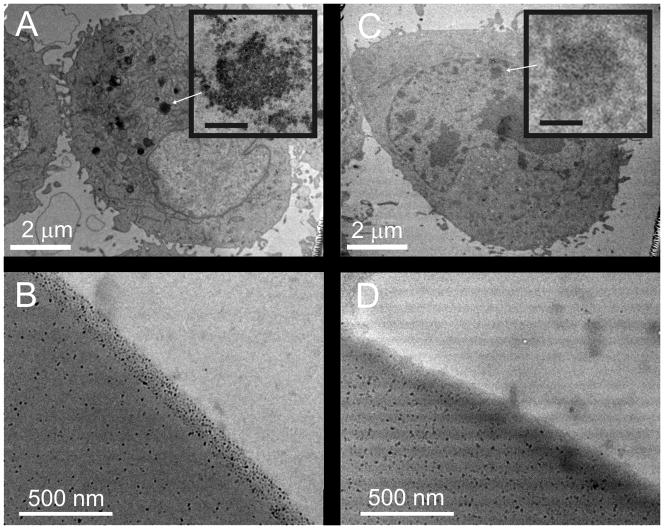
Figure 3.
Uptake of maghemite nanoparticles by cells. TEM images of HeLa cells cultured on 1% magnetic 1002F without (A) and with (C) a 2 μm-thick protective film of native 1002F over the magnetic photoresist. Arrows show clusters of nanoparticles within the cells. Inserts show enlarged images of the magnetic nanoparticles (A) and cellular organelles without nanoparticles (C) (scale bars are 150 nm). TEM images of 1002F photoresist containing 1% maghemite nanoparticles before (B) and after surface roughening (D).

