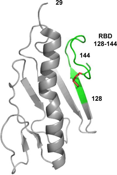
Figure 1.
Ribbon diagram showing the structure of PAK monomeric pilin residues 29–144 (PDB ID: 1DZO). The 17-residue receptor binding domain (RBD) 128–144 is highlighted in green and is defined as one residue (128) prior to the 14-residue disulfide loop and the two residues after the disulfide loop (143 and 144). The disulfide bond between residues 129 and 142 is colored red (12).

