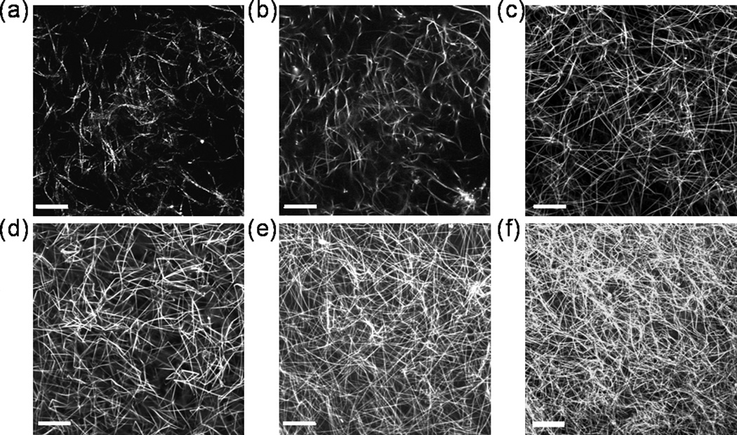
Fig. 1.
Structure of collagen fibers. (a) Reflectance confocal image of 0.3% collagen. (b) Fluorescence confocal image of 0.3% collagen. Fluorescence confocal images of (c) 0.8% (d) 1.0%, (e) 1.5%, and (f) 2.0% collagen. The images in (a) and (b) were of the same sample at the same location. Images are single, horizontal planes in hydrated, TRITC-labeled collagen gels acquired using a 63×, 1.4 NA oil immersion lens with pinhole = 1 Airy unit. Scale bars = 20 microns.

