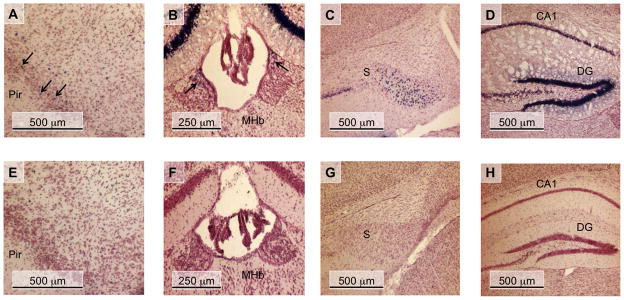
Fig. 3.
nAChR β4 subunit promoter activity of in the CNS of PD30 transgenic mice. Coronal sections of WT transgenic (A - D) and mutant CA box transgenic (E - H) PD30 mouse brains are shown. These sections were simultaneously stained for β-Gal activity, and then counter-stained with neutral red. (A and E) piriform cortex (Pir); (B and F) medial habenula (Mhb); (C and G) subiculum (S); (D and H) hippocampus, corpus ammon layer 1 (CA1), dentate gyrus (DG); Arrows in panels A and B indicate β-Gal-expressing cells.

