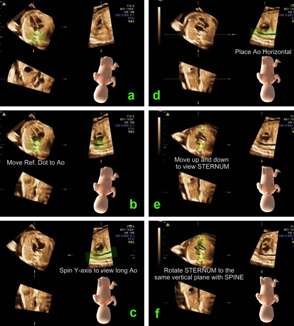
Figure 2.
This image illustrates the steps to standardize virtual fetal position in order to place the fetus in the "exact dorsal supine position". a. In plane A, rotate the spine down to six o'clock. b. In plane A, move the reference dot to the center of the aorta. c. In plane B, spin the volume around the y-axis to display long axis of the descending aorta d. In plane B, rotate the volume around the z-axis to place the descending aorta horizontally. (According to this movement, the volume is placed as if the fetus was lying down on a flat table. However, the fetus may not lie in the exact supine position but more or less on its side.) e. Move to plane A and scrolling parallel up and down through the volume until the sternum echo is depicted. f. Rotate the image in plane A around the z-axis until the sternum and the spine are in a vertical alignment. This last movement results in the exact dorsal supine fetal position.

