
Figure 4.
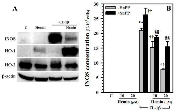
Blockade of hemin-induced iNOS inhibition. A) Culture lysates (40 μg) collected from human astrocytes treated with hemin (20 μM) for 24 h prior to IL-1β (10 ng/ml) exposure for 72 h were electrophorezed, transblotted to nitrocellulose membrane and probed for iNOS, HO-1, HO-2 or β-actin (as internal control) expression. B) Human astrocytes were pretreated with SnPP (10 μM) for 3 h prior to hemin (10 and 20 μM) treatment for 24 h followed by IL-1β (10 ng/ml) exposure for 72 h. Cell lysates were collected, centrifuged and assayed for iNOS expression by ELISA. Sensitivity of iNOS ELISA was 0.05-0.46 U/ml. Data presented are mean ± SE of triplicates from 2 separate experiments using different brain tissue specimens. ** p < 0.01 vs. untreated control (C); ††p < 0.01 vs. IL-1β alone; §§p < 0.01 vs. corresponding hemin+IL-β.