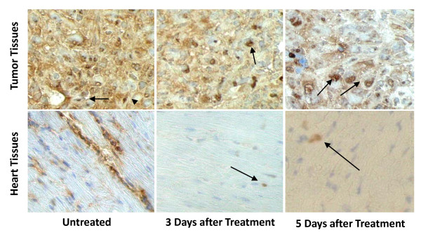
Figure 5.
Effect of doxorubicin on the in vivo expression of B7-H1. Representative Immunohistochemical images (× 540) for B7-H1 (brown) expression in doxorubicin-treated and untreated mice. Shown are sections for tumors formed from xenotransplanted MDA-MB-231 cells in nude mice as well as heart tissues of the nude mice. Nuclei are counterstained with a light hematoxylin to show the nuclear B7-H1 expression. Arrows indicates the nuclear staining of B7-H1.

