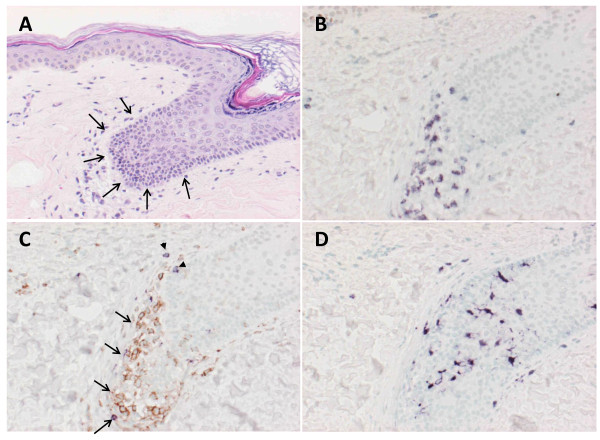
Figure 2.
Immunohistochemical analysis of vitiligo. Biopsy specimens of the area with vitiligo in P2 were stained with H&E to identify infiltrating cells. This focus was a match for vitiligo. To characterize the nature of infiltrating lymphocytes and DCs, biopsy specimens were stained for cell surface markers that were antibodies against CD3, CD4, CD8 and S100. Marked lymphocytes infiltrated into epidermis with depigmentation of biopsy specimen stained with H&E (3A: arrow head). Immunohistochemical analysis revealed that CD3+ T-cells infiltrated at the same sites (3B) and composed of CD4+ T-cells (3C: arrow, brown) and CD8+ T-cells (3C: arrow head, purple). Interestingly, DCs stained with S100 also accumulated at the same sites (3D).

