
Fig. 3.
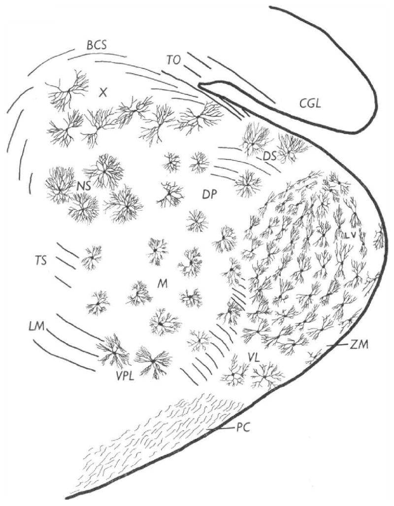
Typical distribution of principal neuron types at the junction of the anterior and middle thirds of the medial geniculate body. Transverse section, Golgi–Cox. 15-day-old cat. (Morest, 1964)
Official websites use .gov
A
.gov website belongs to an official
government organization in the United States.
Secure .gov websites use HTTPS
A lock (
) or https:// means you've safely
connected to the .gov website. Share sensitive
information only on official, secure websites.
Typical distribution of principal neuron types at the junction of the anterior and middle thirds of the medial geniculate body. Transverse section, Golgi–Cox. 15-day-old cat. (Morest, 1964)