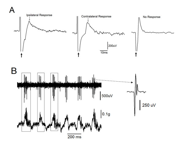
Figure 3.
Identification of spinal motor neurons. A: responses to the gastrocnemius muscle stimulation. The far left and middle panels show the simultaneous responses of spinal motoneurons located ipsilateral and contralateral to the stimulated gastrocnemius muscle, respectively. The right panel shows recordings from the neuron did not respond to muscle stimulation. B: motoneurons were further identified when their spontaneous activity (upper panel) was time locked with spontaneous contractions at the ipsilateral muscle (lower panel).

