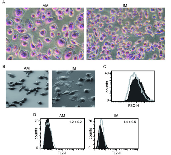
Figure 1.
Morphology and CD90 staining. MΦ visualization by Pappenheim staining (A) and electron microscopy (B). Images are representative for cell preparations from at least two different donors. C: Comparison of MΦ sizes by forward scatter as measured by flow cytometry. Light grey line: IM; filled/dark grey: AM. D: CD90 staining of AM and IM. Filled/dark grey: isotype control; light grey line: antibody staining. MFI values are given within graphs. Data show one representative out of three independent experiments with cells obtained from different donors.

