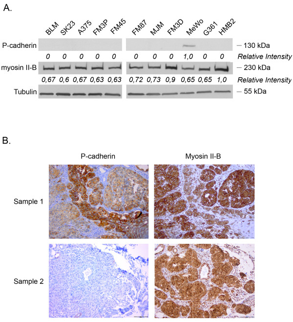
Figure 5.
(A) Western blot to determine the relative amounts of P-cadherin and myosin II-B in a series of melanoma cell lines. High expression levels of myosin II-B for all melanoma cell lines, and absent or low expression levels of P-cadherin could be detected. (B) Immunohistochemical staining of myosin II-B and P-cadherin in nodular melanoma sections (NM). P-cadherin staining shows a honeycomb pattern in normal epidermal layers in contrast to tumor sites. The faint and diffuse staining pattern of the tumor sites indicates loss of cellular cohesion. Myosin II-B staining demonstrates strong positivity at tumor sites, whereas no staining could be observed in normal (epi)dermal layers.

