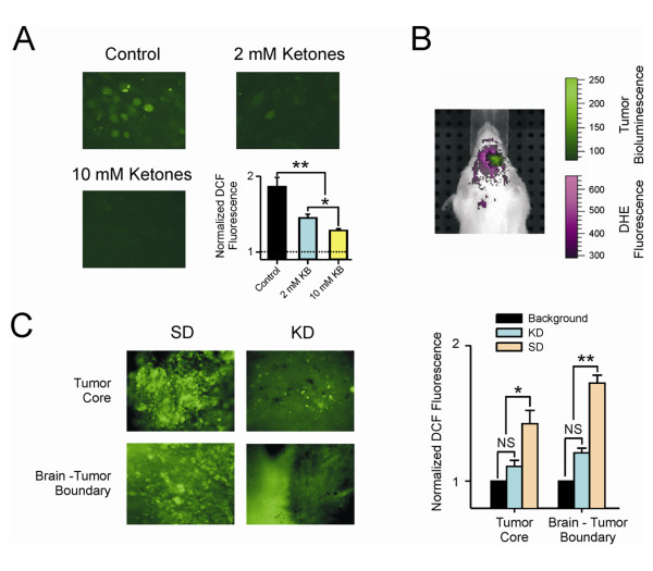
Figure 5.
Ketones reduced ROS levels. (A) Cells were treated with a cocktail containing either 1 mM (total 2 mM ketones) or 5 mM each (total 10 mM ketones) BHB and AcA for 24 hr prior to analysis. ROS levels were analyzed using DCF as described in the methods. Untreated tumor cells had high levels of ROS, and the application of either 2 mM or 10 mM ketones resulted in a dose dependent statistically significant decrease in the DCF signal demonstrating a reduction of ROS. (* = p ≤ 0.05),(* * = p ≤ 0.001); (B) The presence of increased reactive oxygen species (ROS) in tumor and the surrounding area relative to normal tissue is shown in a mouse fed a standard diet. Dihydroethidium (DHE) was imaged using an excitation wavelength of 500 nm and an emission wavelength of 620 nm. Spectral unmixing was used to differentiate the DHE signal from autofluorescence (shown in purple). The tumor was visualized using bioluminescence (shown in green); (C) Photomicrograph of brain slices from animals fed standard diet (SD) or ketogenic diet (KD). Areas from the tumor core and invading front of the tumor are shown. DCF fluorescence was analyzed as described in the methods section. There is a statistically significant difference in the amount of ROS in tumor vs normal brain when animals fed a standard diet were compared to those fed the ketogenic diet. (* = p ≤ 0.05); N.S. = not significant.

