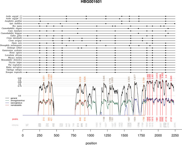
Figure 3.
Example of a graphical representation (stage III) of the multiple nucleotide alignment. This tool was introduced at stage III, to help select conserved regions encompassing introns for PCR primer design. The multiple alignment of the gene family retrieved from Homolens appears at the top; dots indicate intron occurrence (intron positions are reported in gray at the bottom of the graphic). The similarity score ω (black), as well as similarity scores with Strongylocentrotus (ω1, green), Saccoglossus (ω2, blue), and Nematostella (ω3, red) are plotted at the bottom of the graphics; for a better readability, ω1 ... ω3, are halved. Peak of nucleotide conservation and corresponding ω values and positions on the multiple alignment are identified by vertical lines (a colour code indicates the number of species for which additional sequences were available).

